|
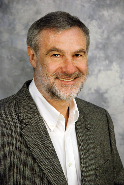
Ernst J. Reichenberger, PhDProfessor, Center for Regenerative Medicine and Skeletal Development, Department of Reconstructive Sciences
|
|||||||||
The skin and skeleton would seem to be very stable parts of the human body. Contrary to appearances, however, both skin and skeleton are in a nearly constant state of flux. The bone mass in skeletal structures is the result of a shifting balance between bone formation and bone resorption, while the skin is constantly sloughed and often subjected to injuries, which must rapidly be repaired. The Reichenberger laboratory is interested in learning about the complex processes required for generating and maintaining the skin and bones. To find out how the mechanisms operate in a healthy person, they study human genetic disorders in which they are disrupted.
In the Reichenberger laboratory, we are interested in dermal and skeletal development and homeostasis. We are currently studying several specific human genetic disorders. For some disorders we have identified disease genes while search for new genes in other disorders. Disease genes interrupt the normal cellular pathways governing developmental and tissue remodeling processes, leading to abnormal tissue behavior.
The selection of disorders like keloid formation, craniometaphyseal dysplasia (CMD), aplasia cutis congenital (ACC) and cherubism (CBM), was guided by our interest in extracellular matrix regulation, skeletogenesis, and bone homeostasis. These diseases can be passed on in autosomal dominant or recessive modes and can also occur sporadically. Collecting affected families and verifying the diagnosis of family members requires a substantial effort, as penetrance and expressivity are variable in all these diseases, and the bone disorders, in particular, are very rare.
Keloids
Injured skin regenerates via a complex wound healing mechanism that leads to scar formation. Keloids are formed when scar tissue does not stop growing but continues to expand over the original margin of the wound like a tumor. In affected families, the tendency to form keloids is an inherited disorder. We use Linkage Analysis and Association Studies (GWAS) to identify keloid genes. Once we have identified the genes responsible for heritable keloid formation and identified the mutations, we will be able to study the functions of these genes and the biological consequences of mutations. Our long-term objective is to use keloid formation as a model to examine the molecular mechanisms leading to neoformation of dermal tissue in fibrotic diseases, as well as in normal wound healing.
Bone Formation Disorders
The goals for the bone projects are to elucidate molecular mechanisms regulating craniofacial bone formation and homeostasis. Maintaining bone mass in every skeletal structure reflects a balance between bone formation by osteoblasts and bone resorption by osteoclasts. Craniometaphyseal dysplasia (CMD) is a rare genetic disorder in which metaphyses of long bones are flared and reveal decreased bone density. Cranial bones show striking overgrowth and increased density of bone. The opposite effect is observed in cherubism (CBM). CBM is a disorder of age-related bone remodeling that is limited to the maxilla and the mandible. During childhood, increased osteoclastogenesis leads to loss of bone in the jaws in symmetrical lesions and replacement of bone with large amounts of fibrous tissue that keeps proliferating like a tumor and can lead to severe swelling of the jaws. Children born with aplasia cutis congenita (ACC) have absent or very thin skin, usually on the scalp. Sometimes the underlying bone is affected as well. ACC can be inherited or appear spontaneously, supposedly by de-novo mutations.
We have recently identified the genes for the autosomal dominant forms of cherubism and CMD.
In cherubism, excessive bone resorption and the characteristic tumor-like growth of proliferating tissue with excessive extracellular matrix deposition are caused by a mutation in a small signal transduction molecule, SH3BP2. In CMD, mutations in the ANKH protein are responsible for the disorder. The transmembrane protein ANKH is known to transport pyrophosphate out of cells and possibly has additional functions. ANKH is a regulator of bone mineralization and possibly prevents mineralization in all other tissues. The ANKH mutations have a profound effect on the function of osteoblasts and osteoclasts but we do not yet understand why this CMD gene mutation affects mainly bones of the face and skull.
Future Directions
Our immediate goal is to study the biological functions of ANK and SH3BP2 on a biochemical and cell biological level. Effects of mutations on upstream and downstream reaction partners of these genes will be investigated and tested in vivo in animal models and in vitro in cell culture and organ culture model systems. For the keloid project as well as for the bone projects we are actively recruiting more patients to participate in the study in order to identify new genes that cause disorders.
Accepting students for Lab Rotations: Spring 1 and 2 Block 2026
Email for more information.
Journal Articles
-
Skeletal abnormalities caused by a Connexin43R239Q mutation in a mouse model for autosomal recessive craniometaphyseal dysplasia.
Bone research 2025 Jan;13(1):14
-
ENPP1 enzyme replacement therapy improves ectopic calcification but does not rescue skeletal phenotype in a mouse model for craniometaphyseal dysplasia.
JBMR plus 2024 Sep;8(9):ziae103
-
Loss-of-function OGFRL1 variants identified in autosomal recessive cherubism families.
JBMR plus 2024 Jun;8(6):ziae050
-
Generation of Keratinocytes from Human Induced Pluripotent Stem Cells Under Defined Culture Conditions.
Cellular reprogramming 2020 Dec;
-
Investigating global gene expression changes in a murine model of cherubism.
Bone 2020 Mar;135115315
-
Clinicoradiologic follow up of cherubism with aggressive characteristics: a series of 3 cases.
Oral surgery, oral medicine, oral pathology and oral radiology 2019 Nov;128(5):e191-e201
-
Alveolar Bone Protection by Targeting the SH3BP2-SYK Axis in Osteoclasts.
Journal of bone and mineral research : the official journal of the American Society for Bone and Mineral Research 2019 Oct;
-
Genetic Disruption of Anoctamin 5 in Mice Replicates Human Gnathodiaphyseal Dysplasia (GDD).
Calcified tissue international 2019 Feb;
-
Rapid degradation of progressive ankylosis protein (ANKH) in craniometaphyseal dysplasia.
Scientific reports 2018 Oct;8(1):15710
-
Second-generation SYK inhibitor entospletinib ameliorates fully established inflammation and bone destruction in the cherubism mouse model.
Journal of bone and mineral research : the official journal of the American Society for Bone and Mineral Research 2018 Apr;331513-1519
-
Rescue of a cherubism bone marrow stromal culture phenotype by reducing TGFβ signaling.
Bone 2018 Mar;11128-35
-
Craniometaphyseal Dysplasia Mutations in ANKH Negatively Affect Human Induced Pluripotent Stem Cell Differentiation into Osteoclasts.
Stem cell reports 2017 Oct;91369-1376
-
Identification of ASAH1 as a susceptibility gene for familial keloids.
European journal of human genetics : EJHG 2017 Oct;25(10):1155-1161
-
Clinicopathologic and Molecular Characteristics of Familial Cherubism with Associated Odontogenic Tumorous Proliferations.
Head and neck pathology 2017 Jul;12136-144
-
Three novel ANO5 missense mutations in Caucasian and Chinese families and sporadic cases with gnathodiaphyseal dysplasia.
Scientific reports 2017 Feb;740935
-
Dietary phosphate supplement does not rescue skeletal phenotype in a mouse model for craniometaphyseal dysplasia.
Journal of negative results in biomedicine 2016 Oct;15(1):18
-
Enhanced TLR-MYD88 signaling stimulates autoinflammation in SH3BP2 cherubism mice and defines the etiology of cherubism.
Cell reports 2014 Sep;8(6):1752-66
-
A site-specific integrated Col2.3GFP reporter identifies osteoblasts within mineralized tissue formed in vivo by human embryonic stem cells.
Stem Cells Translational Medicine 2014 Aug;3(10):1125-37
-
Dental Anomalies Associated with Craniometaphyseal Dysplasia.
Journal of dental research 2014 Mar;93(6):553-558
-
Recruitment of Yoruba families from Nigeria for genetic research: experience from a multisite keloid study.
BMC medical ethics 2014 Jan;15(1):65
-
Induced pluripotent stem cell reprogramming by integration-free Sendai virus vectors from peripheral blood of patients with craniometaphyseal dysplasia.
Cellular reprogramming 2013 Dec;15(6):503-13
-
Mutations in KCTD1 cause scalp-ear-nipple syndrome.
American journal of human genetics 2013 Apr;92(4):621-6
-
Dental abnormalities in a mouse model for craniometaphyseal dysplasia.
Journal of dental research 2013 Feb;92(2):173-9
-
A novel autosomal recessive GJA1 missense mutation linked to Craniometaphyseal dysplasia.
PloS one 2013 Jan;8(8):e73576
-
Oculofaciocardiodental syndrome: a rare case and review of the literature.
The Cleft palate-craniofacial journal : official publication of the American Cleft Palate-Craniofacial Association 2012 Sep;49(5):e55-60
-
Cherubism: best clinical practice.
Orphanet journal of rare diseases 2012 May;7 Suppl 1S6
-
Two novel large ANKH deletion mutations in sporadic cases with craniometaphyseal dysplasia.
Clinical genetics 2012 Jan;81(1):93-5
-
3BP2-deficient mice are osteoporotic with impaired osteoblast and osteoclast functions.
The Journal of clinical investigation 2011 Aug;121(8):3244-57
-
A Phe377del mutation in ANK leads to impaired osteoblastogenesis and osteoclastogenesis in a mouse model for craniometaphyseal dysplasia (CMD).
Human molecular genetics 2011 Mar;20(5):948-61
-
Inhibitory activities of omega-3 Fatty acids and traditional african remedies on keloid fibroblasts.
Wounds : a compendium of clinical research and practice 2011 Jan;23(4):97-106
-
Cherubism gene Sh3bp2 is important for optimal bone formation, osteoblast differentiation, and function.
American journal of orthodontics and dentofacial orthopedics : official publication of the American Association of Orthodontists, its constituent societies, and the American Board of Orthodontics 2010 Aug;138(2):140.e1-140.e11; discussion 140-1
-
Pro416Arg cherubism mutation in Sh3bp2 knock-in mice affects osteoblasts and alters bone mineral and matrix properties.
Bone 2010 May;46(5):1306-1315
-
Introduction of a Phe377del mutation in ANK creates a mouse model for craniometaphyseal dysplasia.
Journal of bone and mineral research : the official journal of the American Society for Bone and Mineral Research 2009 Jul;24(7):1206-15
-
Increased myeloid cell responses to M-CSF and RANKL cause bone loss and inflammation in SH3BP2 "cherubism" mice.
Cell 2007 Jan;128(1):71-83
-
MMP-20 is predominately a tooth-specific enzyme with a deep catalytic pocket that hydrolyzes type V collagen.
Biochemistry 2006 Mar;45(12):3863-74
-
Clinical features of tricho-dento-osseous syndrome and presentation of three new cases: an addition to clinical heterogeneity.
Oral surgery, oral medicine, oral pathology, oral radiology, and endodontics 2005 Dec;100(6):736-42
-
Activation of NFkappaB signal pathways in keloid fibroblasts.
Archives of dermatological research 2004 Aug;296(3):125-33
-
Genome scans provide evidence for keloid susceptibility loci on chromosomes 2q23 and 7p11.
The Journal of investigative dermatology 2004 May;122(5):1126-32
-
Clinical genetics of familial keloids.
Archives of dermatology 2001 Nov;137(11):1429-34
-
Autosomal dominant craniometaphyseal dysplasia is caused by mutations in the transmembrane protein ANK.
American journal of human genetics 2001 Jun;68(6):1321-6
-
Mutations in the gene encoding c-Abl-binding protein SH3BP2 cause cherubism.
Nature genetics 2001 Jun;28(2):125-6
-
Gene for the human transmembrane-type protein tyrosine phosphatase H (PTPRH): genomic structure, fine-mapping and its exclusion as a candidate for Peutz-Jeghers syndrome.
Cytogenetics and cell genetics 2001 Jan;92(3-4):213-6
Letters
-
Noonan-like syndrome mutations in PTPN11 in patients diagnosed with cherubism.
Clinical genetics 2005 Aug;68(2):190-1
Reviews
-
Dental abnormalities in rare genetic bone diseases: Literature review.
Clinical anatomy (New York, N.Y.) 2023 Sep;
-
GeneReviews(®)
1993 Jan;
